Scientists discover new mechanisms of early metastatic spread in breast cancer
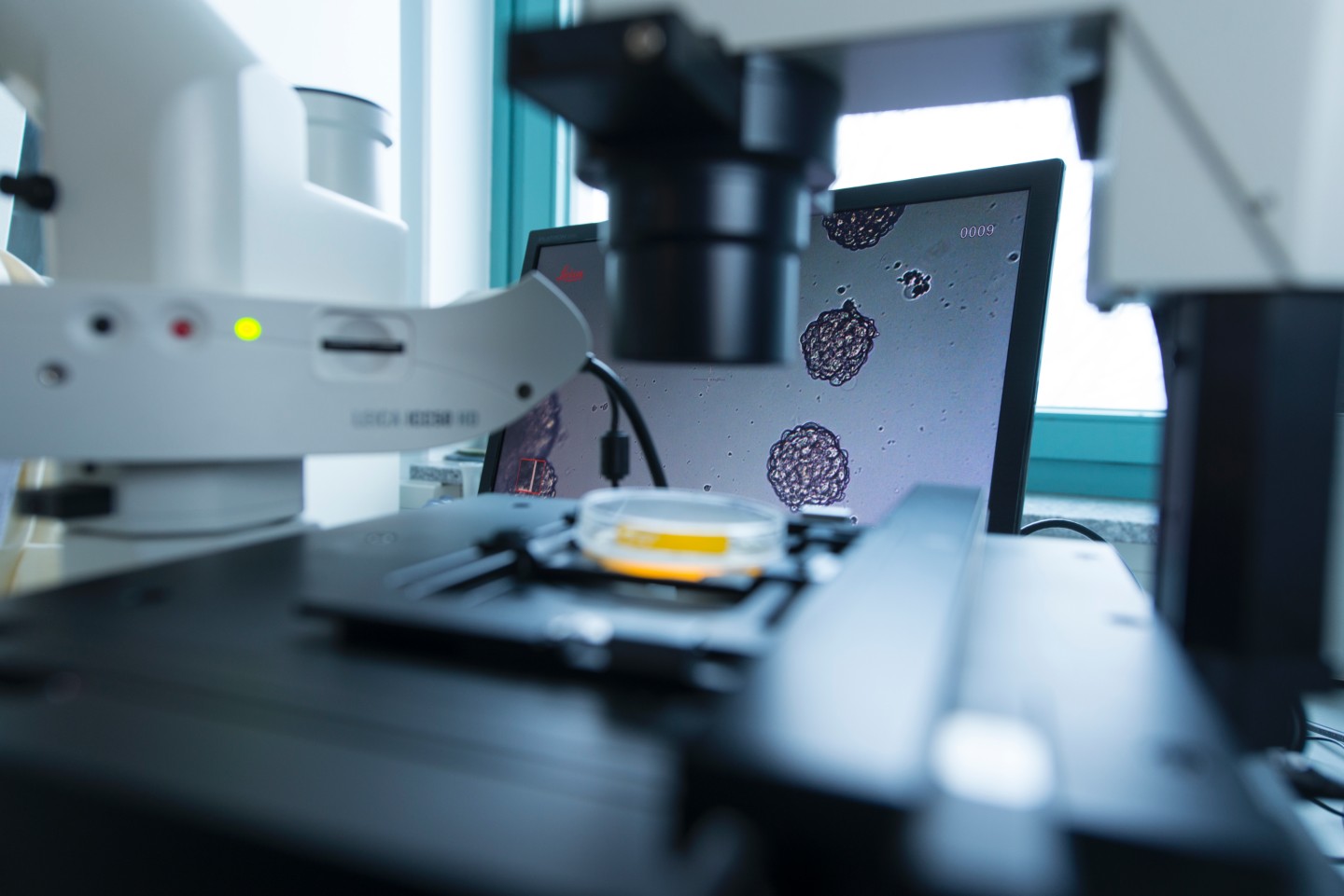
(Hannover/Regensburg, Germany) Scientists of the Regensburg-based Project Group for Personalized Tumor Therapy of the Fraunhofer Institute for Toxicology and Experimental Medicine ITEM and the University of Regensburg and their colleagues from the Icahn School of Medicine at Mount Sinai have discovered new mechanisms of early metastatic spread in breast cancer. Results have been published in the latest issue of the renowned journal Nature (doi:10.1038/nature20785, doi:10.1038/nature20609).
Over several decades, cancer research pursued the dogma that cancer cells seed above all from advanced, i.e. large tumors. This concept was based on the finding that early diagnosis and surgical removal are decisive for cancer patients to be cured. Recently, however, the validity of this concept in explaining treatment success has been questioned increasingly, because patients with small tumors also develop metastases. In addition, disseminated cancer cells often do not show the expected similarity to the primary tumors in their genetic profiles. Disseminated cancer cells, as precursors of metastases, often seem to derive from early stages of primary tumor evolution. Another finding that did not quite confirm the concept of a late-acquired dissemination capacity was the observation that the number of disseminated cancer cells did not correlate with primary tumor size. This spawned the hypothesis that large tumors might be less able to metastasize than small tumors.
Researcher teams under Prof. Christoph Klein in Regensburg and Prof. Julio Aguirre-Ghiso in New York for the first time ever studied mechanisms of early metastatic spread in breast cancer. They found that breast cancer formation hijacks physiological processes controlling mammary epithelial branching and expansion during adolescence and pregnancy, deregulates and then uses them for tumor cell dissemination. Once disseminated, these cells settle in other tissues of the organism and, after further genetic alterations, can grow to often life-threatening metastases in the target organ. The two research teams initially worked independently, but then collaborated and published their results simultaneously in Nature. “Our fundamentally new findings will hopefully advance cancer research substantially,” Klein says.
A few years ago already, the scientists in Regensburg had demonstrated early dissemination of cancer cells. What they now wanted to find out was what mechanisms are activated in early-stage mammary cancer cells and whether and why advanced primary tumors could lose this capacity, once they grow beyond a certain size. They found that the steroid hormone progesterone plays a central role in early lesions, also through indirect effects on other cells. In early stages of cancer development, characterized by low cell density and moderate activation of the oncogene HER2, the progesterone-induced alterations were seen to result in increased migration of cancer cells and acquisition of so-called stemness features, both major prerequisites for metastasis. Interestingly, the same hormone suppresses migration and stemness in advanced primary tumor cells. The decisive factors in reducing the tumor’s dissemination capacity as identified by the Regensburg researchers are increased cell density and strong activation of HER2, which eventually are responsible for increased tumor growth. The switch in the effect of progesterone is regulated by tumor cell density and so-called microRNA, which mediates progesterone receptor downregulation in the cell. The Regensburg researchers furthermore were able to demonstrate that at least 80 percent of the metastases developed in a mouse model were derived from early lesions and that their findings, which they obtained above all from animal models, are also relevant for human metastatic dissemination.
The focus of the New York group for the past few years has been on analyzing p38 signaling – a signaling pathway that might play a central role in tumor dormancy, the clinically latent stage after successful surgical removal of the primary tumor and manifest metastasis. The New York researchers had investigated this signaling pathway in connection with what is referred to as epithelial-mesenchymal transition (EMT), a process allowing stationary epithelial cells in glands such as the mammary gland to acquire mesenchymal characteristics that confer migratory capacity. The scientists detected that early dissemination of mammary cancer cells is regulated by an interactive process between the tumor suppressor gene p38 and the oncogene HER2. Interestingly, p38 signaling had also been identified as a relevant pathway by the Regensburg scientists. During mammary gland development, p38, HER2, and EMT are alternatingly switched on and off. Like the experimental results obtained in Regensburg, the findings of the New York researchers clearly suggested that a physiological process playing a central role in mammary gland development and in the changes occurring during pregnancy is hijacked by tumor cells and used for metastatic dissemination.
The results of the two research teams could also explain why about 5 percent of cancer patients develop metastases, even though no primary tumor can be detected: it is well conceivable that cancer cells migrating from a developing early lesion may be more “successful” somewhere else in the body than the cancer cells remaining at the original site.
The researchers hope that the proposed mechanism will provide a general framework for understanding metastasis formation. According to their findings, cancer cells first go through an early dissemination stage at low cell density at the primary site and later on switch to a proliferation stage when high cell density is reached. However, the Regensburg scientists also found that tumor cells can “relearn” to disseminate even at later stages – from areas of low cell density. This is probably why metastases may derive from different stages of primary tumor evolution, including early and later stages. “Since these stages comprise genetically different cells and modern therapeutic approaches often target genetic alterations, therapies targeting the seed of metastasis need to address this heterogeneity of cells to be successful,” Klein summarizes.