Chip-based cell analysis by chip cytometry

New technology at Fraunhofer ITEM enables sputum analysis by chip cytometry

Chip cytometry enables preservation of the cell material after analyses
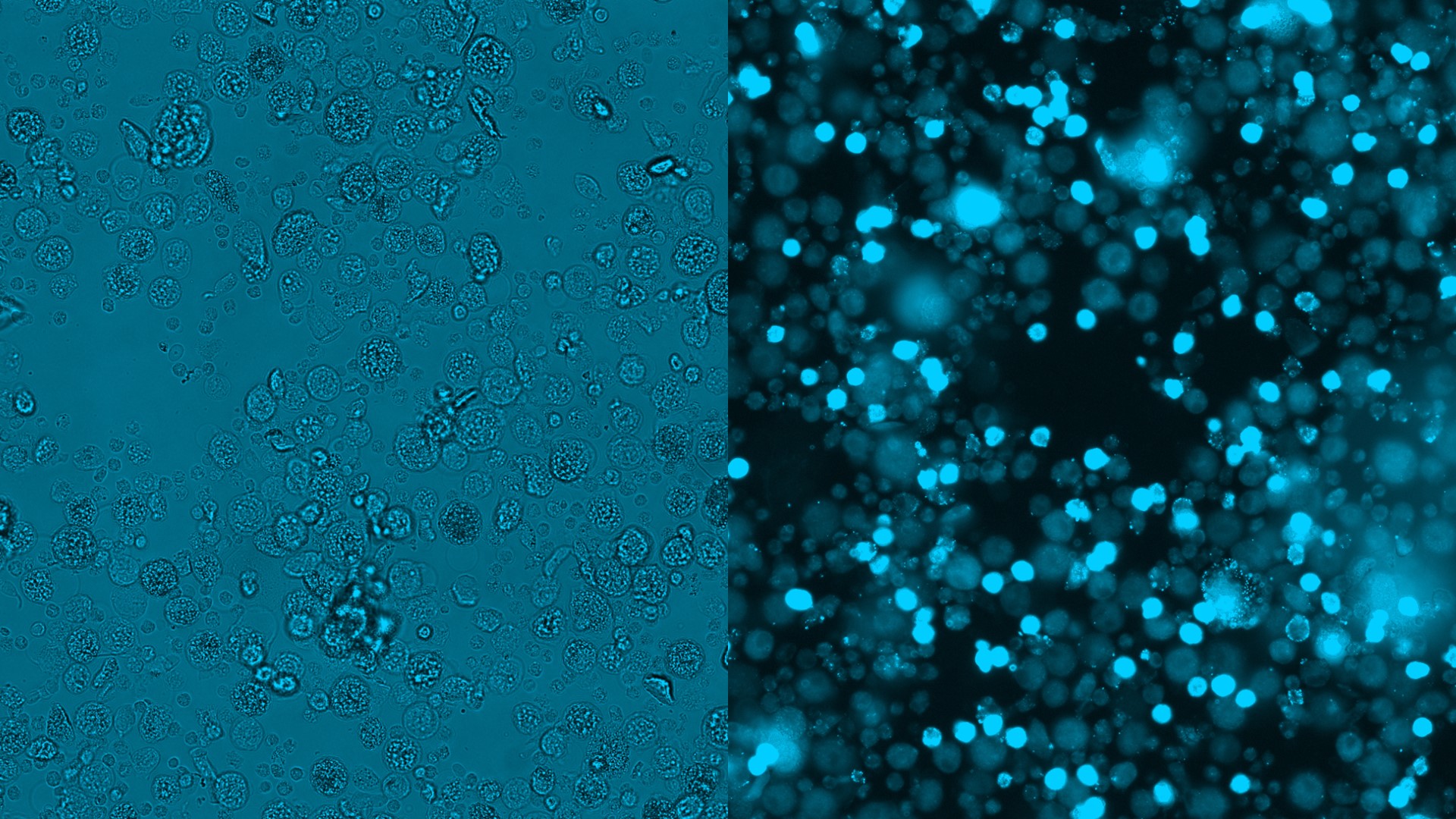
Cell populations within a sputum sample, e.g. macrophages and granulocytes, differ morphologically and biologically. Chip cytometry enables direct comparison of a morphological transmitted-light image and fluorescence staining at the single-cell level.
© Fraunhofer ITEM
Cell populations within a sputum sample, e.g. macrophages and granulocytes, differ morphologically and biologically. Chip cytometry enables direct comparison of a morphological transmitted-light image and fluorescence staining at the single-cell level.

Chip cytometry is based on the immobilization of cells on special object slides referred to as chips. On such chips, cells can be studied with respect to morphology, expression of surface markers, and intracellular function. Consecutive iterative staining processes allow a comprehensive immunological and functional characterization of cells. 

This method enables investigations at the single-cell level and, depending on the cell population, the chips can be stored for a longer period of time. Storage of sputum cell populations on these chips is possible for at least three months. Chip cytometry is thus superior to other methods of analysis such as flow cytometry, offering the possibility to combine direct optical analyses with repeated staining at the single-cell level, but in particular because the cell material of the sample can be preserved after the measurements.

Fraunhofer ITEM is validating chip cytometry for induced sputum

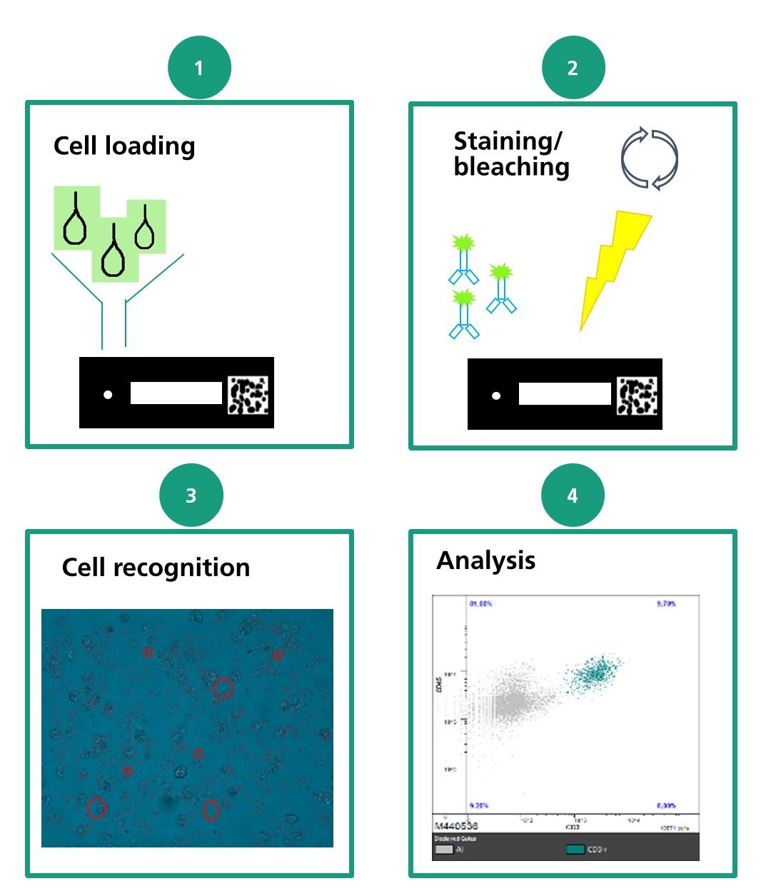
Chip cytometry consists of 4 steps: (1) cell loading, (2) staining/bleaching, (3) cell recognition, and (4) analysis.
© Fraunhofer ITEM
Chip cytometry consists of 4 steps: (1) cell loading, (2) staining/bleaching, (3) cell recognition, and (4) analysis.

Induced sputum obtained from the lungs of test subjects often exhibits limited cell numbers, so that the use of chip cytometry is holding promise for advances in immunological characterization and cell diagnostics. Furthermore, sputum includes cells of different sizes with specific fluorescence properties, placing special requirements on the measurement method used. First data showed good comparability with the standard methods flow cytometry and microscopic cell differentiation.

“Using chip cytometry for a complex matrix such as sputum finally allows cell properties to be studied at the single-cell level. This has the great advantage of making matrices with small numbers of cells accessible to clinical research; and of enabling detailed investigation of these cells without the need to define the endpoints to be investigated prior to study start,” says Dr. Meike Müller, head of the Department of Biomarker Analysis and Development.

Advantages of chip cytometry over other methods

  • Iterative fluorescence staining: the very same sample can be repeatedly stained with sequential antibodies and measured at different time points
  • Preservation of the cell material: sputum samples on chips can be stored for at least three months after measurement of the first biomarker of interest
  • Storage of peripheral blood cell samples on chips for more than 24 months
  • Sample storage on the chip allows measurement of additional endpoints even after study completion (iterative decision making)
  • Exclusion of false-positive events by direct comparison of fluorescence and transmitted-light images
  • Unlimited number of biomarkers can be analyzed in a single tissue or cell suspension sample
  • Preparation and preservation of cells on chips and their storage at different test sites and subsequent centralized analysis without significant degradation of biomarkers allows multicenter use of this method
  • In-depth analysis of single cells

Chip cytometry: "We can do iterative immunophenotyping of cells – even after storage!"

Chip cytometry is based on the immobilization of cells on special object slides, referred to as chips. On these chips, cells can be analyzed with regard to morphology, expression of surface markers, and intracellular function.

Iterative staining protocols allow comprehensive immunological and functional characterization of cells

Privacy warning

With the click on the play button an external video from www.youtube.com is loaded and started. Your data is possible transferred and stored to third party. Do not start the video if you disagree. Find more about the youtube privacy statement under the following link: https://policies.google.com/privacy

Publications

Carstensen, S., Benediktus, E., Litzenburger, T., Hohlfeld, J. M., Muller, M. (2021).
Basophil activation test: Assay precision and BI 1002494 SYK inhibition in healthy and mild asthmatics.
Cytometry A [Epub ahead of print]. doi: 10.1002/cyto.a.24338

Carstensen, S., Holz, O., Hohlfeld, J. M., Muller, M. (2021).
Quantitative analysis of endotoxin-induced inflammation in human lung cells by Chipcytometry.
Cytometry A [Epub ahead of print]. doi: 10.1002/cyto.a.24352

Carstensen S, Holz O, Hohlfeld JM, Müller M (2019). 
Eosinophil and Neutrophil Granulocyte Immunophenotyping Using Chipcytometry 
American Journal of Respiratory and Critical Care Medicine Vol. 199: Abstract A2956

Carstensen S, Holz O, Müller M, Hohlfeld JM (2018).
Validation of cell analysis by chipcytometry in induced sputum cells
American Journal of Respiratory and Critical Care Medicine Vol. 197: Abstract A4768

Contact

Meike Müller

Contact Press / Media

Dr. Meike Müller

Head of Department of Biomarker Analytics and Development

Phone +49 511 5350-8144